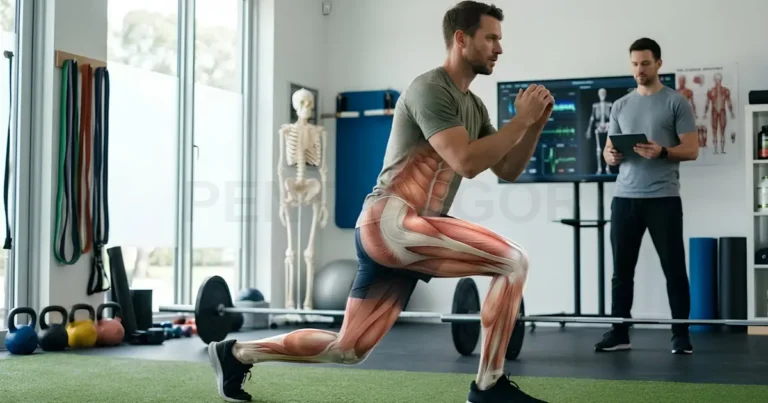
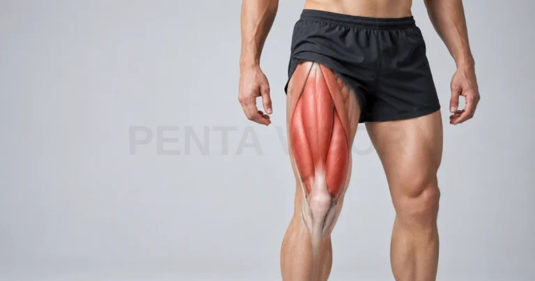
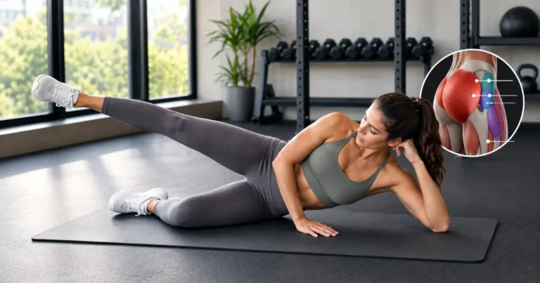
Have you ever wondered how muscle anatomy explains everything from a steady heartbeat to a single step up the stairs? Your muscular system includes more than 600 named skeletal muscles, and it shapes how you move, breathe and age. In this guide, we explain the three muscle types, the major muscle groups, how movement works and which changes are worth discussing with your doctor.
What the Muscular System Is and Why It Matters for Human Movement
The muscular system is the organ system that produces force, movement and heat in the human body. As a muscle organ system, it includes skeletal, cardiac and smooth muscle, all built from contractile tissue that shortens when the nervous system sends a signal. Every action depends on it, from blinking and breathing to climbing stairs, so learning muscle anatomy gives you a practical map of how your body works.
Beyond movement, this system supports posture, protects internal organs, stabilizes joints and helps regulate body temperature. Skeletal muscle is also a major site of glucose uptake and storage, which links muscle health to metabolic health. The next three subsections define the system, its connective tissue partners and its boundary with the skeleton.
Defining the Muscular System as an Organ System
An organ system is a group of organs that work together toward a shared function, and the shared function of the muscular system is contraction. Each individual muscle is an organ made of muscle tissue, connective tissue, blood vessels and nerves. Because muscles carry out commands from the brain and spinal cord, physiologists describe them as effector organs of the nervous system.
This teamwork is clearest at the junction between nerve and muscle. A motor neuron releases a chemical messenger, muscle fibers respond by shortening, and the result is force that moves a bone or narrows a tube. When you lift a cup, signals reach the right arm muscles within a fraction of a second.
How Muscle, Fascia and Tendon Work as One Contractile Unit
A muscle rarely works alone, because fascia and tendon carry its force to the skeleton. Fascia is the connective tissue that wraps and separates muscles, a tendon is the strong cord that joins muscle to bone, and an aponeurosis is a flat, sheet-like tendon, as seen in the abdominal wall. Together they form what many clinicians call a myofascial unit.
Definitions of fascia are still debated among anatomists. The Fascia Nomenclature Committee describes a fascia as a sheath or sheet of dissectible connective tissue that attaches, encloses and separates muscles and organs. The practical point is that force travels through connective tissue as well as muscle fibers, so stiffness or injury can involve either part.
Muscular System vs Musculoskeletal System: Where the Boundary Lies
The muscular system centers on muscles, while the musculoskeletal system is the broader group that also includes bones, joints, cartilage, ligaments and tendons. Think of the muscular system as the engine and the musculoskeletal system as the whole machine, including its frame and hinges. Tendons sit on the boundary, because they act as part of the muscle unit yet are usually classed as connective tissue.
This distinction helps when you read about symptoms. A sprained ankle involves ligaments, a stress fracture involves bone and a strained hamstring involves muscle, yet all three are called musculoskeletal injuries. Next, we look at the three types of muscle that make up the muscular system itself.
The Three Types of Muscle in the Human Body
The human body contains three types of muscle: skeletal, cardiac and smooth. These muscle types in the human body differ in how they look under a microscope, in how the nervous system controls them and in where they sit.
Below is a detailed breakdown of the unique characteristics defining each muscle type.
Skeletal Muscle: Voluntary Movement and Posture
Skeletal muscle is striated muscle that you can move on purpose, and it is the type that drives posture and voluntary movement. Its long fibers contain repeating units that create a striped look under a microscope, and tendons anchor it to bone. Even while you sit still, small postural muscles work constantly, mostly without conscious effort.
Skeletal muscle is what most people mean by “muscles” in everyday conversation. When you walk, type or hold your head upright, you rely on it. Because it responds to training, it is also the muscle type that changes most with exercise, inactivity and age.
Cardiac Muscle: The Myocardium and Involuntary Rhythm
Cardiac muscle is the involuntary, striated muscle of the heart wall, known as the myocardium. Its cells branch and connect end to end through intercalated discs, which let electrical signals pass quickly from cell to cell. This design allows the heart to contract as one coordinated unit and keep a steady rhythm without conscious control. Because the myocardium never rests for long, it depends on a constant blood supply.
Smooth Muscle: Visceral Control in Hollow Organs
Smooth muscle is non-striated, involuntary muscle found in the walls of hollow organs and blood vessels. It moves food along the intestines, narrows or widens blood vessels, empties the bladder and adjusts airway diameter. The autonomic nervous system and hormones control it, so you do not direct it consciously. Because it surrounds internal organs, it is also called visceral muscle.
Smooth muscle contracts more slowly than skeletal muscle but can hold tension for long periods with little energy. For example, the smooth muscle in your artery walls helps keep blood pressure in a healthy range throughout the day. With the three types in mind, we can now ask how many muscles the human body actually contains.
How Many Muscles Are in the Human Body? What the Evidence Says
Most anatomy references list roughly 600 to 650 named skeletal muscles in the human body, but no single number is universally accepted. The count applies to skeletal muscle, because cardiac and smooth muscle are tissues made of enormous numbers of cells rather than separate named units. Together, skeletal muscles make up roughly 30 to 40 percent of body mass.
If you have seen figures such as 640, 650 or even higher, you have not misread anything. The difference reflects how each source defines and divides a muscle, not a disagreement about the body itself. The next subsection explains where those differences come from.
Why Anatomists Disagree on the 600 to 850 Muscle Count
Anatomists disagree on the count because there is no single rule for where one muscle ends and the next begins. Some references count the four parts of the quadriceps as separate muscles, while others treat the group as one. Multi-headed muscles such as the biceps brachii, and layers that blend together along the spine, are also counted differently. Counts that split muscles into fine subdivisions are higher, and figures up to about 850 appear in some sources.
Natural variation adds another layer. The palmaris longus, a slender forearm muscle, is absent in roughly 10 to 15 percent of people, usually without any noticeable effect on function. A muscle count is therefore best read as a well-informed estimate, not a fixed inventory of the body.
Terminologia Anatomica and the Standard European Naming Convention
Terminologia Anatomica is the international standard list of anatomical terms, published with Latin names and English equivalents. Its second edition (TA2) was released online in 2019 by the Federative International Programme for Anatomical Terminology (FIPAT) and adopted by the International Federation of Associations of Anatomists in 2020. Standard Latin terms, such as musculus gluteus maximus, let clinicians across Europe and beyond describe the same structure in the same way.
In practice, this matters to students in many European medical schools, where Latin terms appear in lectures, imaging reports and exams. In our articles, we give the common English name first and add the standard Latin term where it helps you cross-check other sources. Next, we move from counting muscles to locating them, beginning with the front of the body.
Anterior Muscle Map: Front-of-Body Muscles at a Glance
The anterior muscle map shows the muscles on the front of the body, from the face to the feet. It is the view used most often in human muscle anatomy charts because so many large, familiar muscles sit here. Use this section for orientation, and turn to our regional guides for origins, insertions and nerve supply.
Front Muscles of the Head, Neck and Torso
The front of the head, neck and torso holds muscles for facial expression, chewing, head movement, breathing and trunk flexion. You can feel several of them on your own body, which makes this region a good starting point for learning. The checklist below names the main ones you will meet on an anterior chart.
- Face and jaw: orbicularis oculi, orbicularis oris, masseter and temporalis
- Front of the neck: sternocleidomastoid, platysma and the infrahyoid (strap) muscles
- Chest: pectoralis major, pectoralis minor and serratus anterior
- Abdominal wall: rectus abdominis, external and internal obliques and transversus abdominis
- Breathing: intercostal muscles and the diaphragm, which lie deep and are not visible on the surface
For example, when you clench your teeth, you can feel the masseter bulge at the angle of your jaw. Deeper muscles such as the transversus abdominis, which works like a natural corset, do not show on the surface but stabilize your trunk. Each area has its own detailed guide in our regional series.
Front Muscles of the Upper and Lower Limbs
On the front of the limbs, the main muscles bend the elbow, extend the knee and lift the foot. In the arm, the front muscles are mostly flexors, while in the thigh they are mainly extensors of the knee. The checklist below groups them by region, from the shoulder to the ankle.
- Shoulder and upper arm: anterior deltoid, biceps brachii, brachialis and coracobrachialis
- Forearm: wrist and finger flexors, pronator teres and brachioradialis
- Hip and thigh: iliopsoas, sartorius, quadriceps femoris and the adductor group
- Lower leg: tibialis anterior, extensor digitorum longus and extensor hallucis longus
The quadriceps femoris is a group of four muscles that share one tendon at the kneecap. When you stand up from a chair, it works together with the muscles at the back of your hip. The rear view completes the picture, so we turn to it next.
Posterior Muscle Map: Back-of-Body Muscles at a Glance
The posterior muscle map shows the muscles on the back of the body, many of which keep you upright and power walking, lifting and pushing off the ground. These muscles often work against gravity, so they can feel strained after long hours of sitting. As with the front view, this section is a summary, and our regional guides cover each muscle in detail.
Rear Muscles of the Trunk and Shoulder Girdle
The rear muscles of the trunk and shoulder girdle hold the shoulder blade in position and extend, rotate and stabilize the spine. Some of them, such as the trapezius, are visible on the surface, while others lie in deeper layers close to the spine. The checklist below lists the main groups from the surface to the deeper layers.
- Superficial back: trapezius and latissimus dorsi
- Shoulder blade stabilizers: rhomboid major, rhomboid minor and levator scapulae
- Rotator cuff: supraspinatus, infraspinatus, teres minor and the deeper subscapularis
- Spinal extensors: erector spinae group and multifidus
- Shoulder cap: posterior deltoid
The trapezius is a large, diamond-shaped muscle that spans the neck and mid-back. Its upper fibers lift the shoulder blade when you shrug, while its lower fibers help draw it down and back. Because many back muscles overlap, anatomists usually describe them in layers, from superficial to deep.
Rear Muscles of the Arm, Hip and Leg
The rear muscles of the arm, hip and leg straighten the elbow, extend the hip and knee, and push the foot downward. They lengthen the body when you stand tall and propel you forward when you walk. The checklist below shows the main muscles by region.
- Upper arm: triceps brachii
- Forearm: wrist and finger extensors
- Hip: gluteus maximus, gluteus medius, gluteus minimus and deep rotators such as the piriformis
- Thigh: hamstrings (biceps femoris, semitendinosus and semimembranosus)
- Calf: gastrocnemius, soleus and the Achilles tendon
The gluteus maximus is the largest muscle in the body by mass, and it drives hip extension when you climb stairs. The gastrocnemius and soleus together form the triceps surae, which pushes you off the ground with every step. With both maps in view, we can now sort these muscles into functional groups.
The 12 Major Muscle Groups and What Each One Does
The 12 major muscle groups are a teaching framework that sorts the body’s largest muscles by region and function. They are the chest, back, shoulders, upper arms, forearms, abdominal wall, obliques, spinal extensors, glutes, quadriceps, hamstrings and calves. No official body fixes the number at 12, and different sources list different totals, but this version is common in education and covers most large muscles.
Upper Body Groups: Chest, Back, Shoulders and Arms
The upper body groups are the chest, back, shoulders and arms, and together they push, pull, lift and position your hands. We count the arms as two groups, upper arms and forearms, which brings the upper body to five groups.
- Chest (pectoralis major and minor): pushes and draws the arm across the body
- Back (latissimus dorsi, trapezius and rhomboids): pulls the arm down and back and controls the shoulder blade
- Shoulders (deltoid and rotator cuff): raise and rotate the arm
- Upper arms (biceps brachii, brachialis and triceps brachii): bend and straighten the elbow
- Forearms (wrist and finger flexors and extensors): move the wrist and fingers and control grip
When you pull open a heavy door, your back and biceps pull, your shoulder stabilizers hold the joint steady and your forearm muscles maintain the grip. This is a typical example of groups working in sequence rather than alone. Most everyday tasks use several groups at once.
Core Groups: Abdominal Wall, Obliques and Spinal Extensors
The core groups are the abdominal wall, the obliques and the spinal extensors, and they stabilize the trunk between the ribs and the pelvis. Unlike the limb muscles, they mostly work to hold the body steady rather than to create large movements. Each group has a distinct job, as the checklist below shows.
- Abdominal wall (rectus abdominis and transversus abdominis): flexes the trunk and compresses the abdomen
- Obliques (external and internal): rotate and side-bend the trunk
- Spinal extensors (erector spinae and multifidus): extend the spine and keep you upright
Anatomists often compare the core to a cylinder, with the diaphragm on top and the pelvic floor below. When you carry a shopping bag on one side, your obliques and deep spinal muscles resist the pull so that you stay upright. Coordination and endurance matter as much as size in this region.
Lower Body Groups: Glutes, Quadriceps, Hamstrings and Calves
The lower body groups are the glutes, quadriceps, hamstrings and calves, and they carry your weight, propel walking and absorb impact. Each group works mainly on a different joint, the hip, the knee or the ankle. The checklist below shows what each one does in daily movement.
- Glutes (gluteus maximus, medius and minimus): extend and stabilize the hip
- Quadriceps (rectus femoris and the three vasti): extend the knee
- Hamstrings (biceps femoris, semitendinosus and semimembranosus): bend the knee and extend the hip
- Calves (gastrocnemius and soleus): lift the heel and push off
When you climb a flight of stairs, your glutes and quadriceps lift your body, your hamstrings control the knee and your calves add the final push. Because these groups are so large, they matter greatly for mobility as you age. The table below brings all 12 groups together.
A Quick-Reference Guide to the 12 Major Muscle Groups
Here is a quick-reference guide detailing the 12 major muscle groups, including where they are located and how they function in the body. You can use this overview as a helpful checklist or study aid when reviewing anatomical diagrams. Please note that the neck muscles, hip adductors, and shin muscles fall outside this general framework and are typically covered in specialized regional anatomy guides.
- Chest: Located on the front of the upper torso, these muscles are primarily responsible for pushing movements and adducting the arms toward the center of the body.
- Back: Spanning the upper and mid-back as well as the sides of the trunk, this large group pulls the arms backward and downward while controlling the movement of the shoulder blades.
- Shoulders: Sitting like a protective cap over the shoulder joints, these muscles function to raise and rotate the arms.
- Upper arms: Positioned on both the front and back of the upper arm, these muscles allow you to bend and straighten your elbows.
- Forearms: Located between the elbow and the wrist, they control your grip strength and drive the fine movements of your wrists and fingers.
- Abdominal wall: Found on the front of the trunk, the abs work to compress the abdomen and flex the torso forward.
- Obliques: Running along the sides of the trunk, these muscles enable torso rotation and side-bending.
- Spinal extensors: Stretching along the length of the spine, they are crucial for extending the back and maintaining an upright post.
- Glutes: Comprising the buttocks and the outer sides of the hips, this powerful group extends, abducts, and rotates the hip joints.
- Quadriceps: Covering the front of the thighs, their primary mechanical job is to extend and straighten the knees.
- Hamstrings: Located on the back of the thighs, they function to bend the knees and extend the hips backward.
- Calves: Found on the back of the lower legs, these muscles lift the heels and provide the necessary push-off power for walking, running, and jumping.
Understanding the location of each muscle group is only the first step. The second crucial component is learning exactly how these muscles pull against your bones to generate motion whether you are standing perfectly still, stepping down from a curb, or simply lifting a cup. In the following section, we will break down the underlying mechanics of skeletal muscle movement.
How Skeletal Muscles Produce Movement
Skeletal muscles produce movement by contracting and pulling on bones through tendons, which turns a joint like a hinge or a pivot. A muscle can only pull, never push, so the body uses opposing muscles to create motion in both directions. The next three subsections explain attachments, muscle roles and leverage.
Origin, Insertion and Line of Pull Explained
A muscle’s origin is the attachment that usually stays more fixed, its insertion is the attachment that usually moves, and its line of pull is the direction in which it applies force. Origins are typically closer to the trunk, and insertions are typically farther out on the limb. The biceps brachii, for example, originates on the scapula and inserts on the radius, so its contraction bends the elbow.
These terms are conventions, and roles can reverse. During a chin-up, your hands are fixed, so the insertion stays still while the origin moves toward it. The line of pull explains why each muscle produces a specific action: fibers running in a different direction would pull the bone along a different path.
Agonist, Antagonist, Synergist and Fixator Roles
Muscles work in teams, and each muscle in the team plays one of four roles during a movement. These roles are not fixed labels, because the same muscle can change roles from one movement to the next. The checklist below defines them.
- Agonist (prime mover): produces the main movement
- Antagonist: opposes or controls the movement
- Synergist: assists the agonist or refines the direction of movement
- Fixator: stabilizes a joint or bone so the movement can happen
During a biceps curl, the biceps brachii is the agonist, the triceps brachii is the antagonist, the brachialis assists as a synergist and the muscles around the shoulder blade act as fixators. Roles change with the task: when you straighten your arm, the triceps becomes the agonist. This is why muscle pairs, such as the quadriceps and hamstrings, are often trained and rehabilitated together.
Lever Systems and Why Some Muscles Generate More Force
A lever system is a rigid bone that turns around a joint, with muscle providing the effort and body weight or an external object providing the load. Anatomists describe three classes, based on where the fulcrum, effort and load sit.
- First class: the fulcrum lies between effort and load, such as nodding the head at the top of the spine
- Second class: the load lies between fulcrum and effort, such as rising onto your toes
- Third class: the effort lies between fulcrum and load, such as bending the elbow to lift a cup
Third-class levers are the most common in the limbs. They favor speed and range of motion, but the muscle must produce more force than the load it lifts. Mechanical advantage also explains why small differences in where a tendon attaches can change how much torque a muscle produces. Next, we compare how these systems look in men and women.
Male and Female Muscular Anatomy: Real Differences and Common Myths
Men and women have the same named muscles, and the real differences lie in average size, distribution and hormonal influence rather than in structure. Many popular beliefs about female muscle anatomy exaggerate these differences or ignore the large overlap between individuals. The two subsections below separate the evidence from the myths.
Distribution, Cross-Sectional Area and Hormonal Influence
In a whole-body MRI study of 468 adults aged 18 to 88 years, men had on average 33.0 kg of skeletal muscle and women had 21.0 kg, which equals 38.4% and 30.6% of body mass. The gap was larger in the upper body, where women had about 40% less muscle than men, than in the lower body, where the gap was about 33%.
Testosterone, which is higher on average in men, promotes muscle protein synthesis and larger muscle fibers. Estrogen appears to influence muscle repair and function, although research is still developing. Strength differences follow a similar pattern, about 30% in the lower body and about 50% in the upper body according to studies cited in the same paper, and individual variation is large. Because of that variation, several myths persist, and the checklist below sets each one against the evidence.
- Myth: Women’s muscles work differently. Fact: The muscles and their actions are the same, and only average size and distribution differ.
- Myth: Strength training makes women bulky. Fact: Most women gain strength steadily and size gradually, because building large muscle typically requires years of focused training.
- Myth: Muscle turns into fat when you stop training. Fact: Muscle and fat are different tissues, so inactivity shrinks muscle fibers while fat can accumulate separately.
Why Labeled Diagrams Often Default to a Male Model
Labeled anatomy diagrams often default to a male body because the male figure has long served as the standard reference in anatomy books and atlases. A 2024 analysis of 11 widely used atlases and textbooks published from 2015 to 2020 found that, among images that could be sorted by sex, 61.6% showed males and 38.4% showed females. The male bias appeared across whole-body and regional images.
European curricula show a similar pattern. A recent analysis of atlases recommended in Polish medical programs found that male bodies appear more often in general anatomy, while female bodies appear mainly in sex-specific illustrations. Use the checklist below to read any diagram critically.
- Check whether the figure states which body it shows
- Compare the same muscle in at least two sources
- Remember that size, shape and attachments vary between individuals
- Use labels to learn names and locations, not as a guide to how your own muscles should look
Recognizing this bias helps you study more accurately and see anatomy as a spectrum, not a single template. It may also help future clinicians recognize normal variation in every patient. Next, we look at what happens to the muscular system as you age.
Muscular System Health Across the European Lifespan
Muscle health changes across the lifespan: strength builds through early adulthood, plateaus and then gradually declines unless you keep your muscles active. A German cohort of more than 200,000 adults found that grip strength rose in the third and fourth decades of life and declined afterward. In the EU, where 22.0% of the population was aged 65 or older on 1 January 2025, this pattern has real public health weight.
Sarcopenia and Age-Related Muscle Loss in EU Populations
Sarcopenia is a progressive, generalized skeletal muscle disorder that is associated with falls, fractures, physical disability and mortality. The European Working Group on Sarcopenia in Older People (EWGSOP2) updated its definition in 2018 and made low muscle strength the first sign. Low strength suggests probable sarcopenia, low muscle quantity or quality confirms it, and poor physical performance marks it as severe.
The checklist below shows the EWGSOP2 thresholds that clinicians commonly use in Europe. They are guides for professionals, not a tool for self-diagnosis, because a diagnosis also depends on your medical history, a physical examination and other tests, such as a body composition scan.
- Low grip strength: under 27 kg for men and under 16 kg for women
- Slow chair stand: more than 15 seconds to rise five times
- Low appendicular muscle mass: under 20 kg for men and under 15 kg for women
- Low muscle mass relative to height: under 7.0 kg per square meter for men and under 5.5 for women
- Poor physical performance: walking speed of 0.8 meters per second or slower
These thresholds come mainly from British data, and European research shows why they are treated as guides. A German study of more than 200,000 adults suggested slightly higher grip cut-offs of 29 kg for men and 18 kg for women. In a multinational trial of adults aged 70 and older in Germany, France, Portugal and Switzerland, prevalence ranged from 0.7% to 16.8% depending on which of twelve definitions was used.
Aging is not evenly spread across Europe. Eurostat reports that 99 million EU residents were aged 65 or older in 2025, with the highest shares in Italy (24.7%), Portugal (24.3%) and Bulgaria (24.0%). Regular muscle-strengthening activity supports muscle health at every age, and WHO recommends it on two or more days a week for adults, including older adults.
For older adults, WHO also advises varied activity that emphasizes balance and strength on three or more days a week, which helps prevent falls. If you have a health condition, speak with your doctor before starting a new program, and choose activities that match your abilities.
Everyday Signals of Muscle Weakness Worth Discussing With a GP
Everyday signals of muscle weakness include trouble rising from a chair, a slower walking pace, a weaker grip and more frequent stumbles. True weakness differs from tiredness, because you cannot produce your usual force even when you try. In many European health systems, your general practitioner (GP) or family doctor is the right first contact. The checklist below can help you notice changes worth mentioning.
- Trouble rising from a chair without pushing with your hands
- A slower walking pace, or needing more rests than before
- A weaker grip, such as difficulty opening jars or carrying bags
- More effort to climb stairs
- Falls or near-falls
- Visible thinning of a limb, or unintentional weight loss
- Weakness that started gradually, is slowly getting worse or comes and goes
Some medicines, alcohol and certain illnesses can also cause muscle weakness. Package leaflets for medicines authorized in the EU list known side effects, including muscle-related ones where relevant, so check the leaflet and talk with your doctor or pharmacist before you stop or change any treatment.
Some signs need urgent care instead of a routine appointment, because weakness with these features can worsen quickly. Call your local emergency number (112 across the EU, 999 in the UK) if weakness comes with any of the signs in the checklist below.
- Weakness that starts over a few days or faster, or that affects one side of the body suddenly
- Trouble breathing
- Trouble chewing, talking or swallowing
- Trouble raising your head while lying down
- Being unable to walk
This guide is educational and does not replace personal medical advice. Once you know how your muscular system changes over time, the next step is learning how to explore each region in detail, which we cover in the next section.
How to Use This Muscular System Hub: Your Guide to Every Region
This hub is a pillar guide: it presents the whole muscular system in one place and points you to detailed articles for each body region. You can read it from top to bottom or jump to the section on any part of human muscle anatomy that answers your question. The suggestions below help you plan your study.
Region-by-Region Reading Path for Students and Clinicians
A region-by-region reading path moves from the head and neck downward, the same order in which most anatomy courses teach the body. This order keeps related muscles together and lets you build on what you already know. Follow the steps below in order, or start with the region you need most.
- Read this guide first to learn the vocabulary and the three muscle types
- Head and neck muscles
- Shoulder girdle and upper back muscles
- Chest, arm and forearm muscles
- Trunk and core muscles
- Hip and glute muscles
- Thigh muscles
- Lower leg and foot muscles
Students can add Latin names and a note on origin, insertion, action and nerve supply for every muscle. Clinicians and therapists may prefer to start with the region they treat most often and return to this hub for whole-body context.
Study Tools, Labeled Charts and Revision Resources
Effective muscle study combines labeled charts, active recall and hands-on surface anatomy. Start with a drawing of the muscular system for orientation, then move to more detailed atlases once you know the main names. The checklist below turns this into a routine.
- Cover the labels on an anterior and a posterior chart, then name each muscle from memory
- Build a table of origin, insertion, action and nerve supply for each muscle
- Learn the Latin term next to the English name
- Locate muscles on your own body, for example the biceps brachii or the quadriceps
- Revise in short sessions over several weeks with spaced-repetition flashcards
- Compare more than one atlas, including at least one that shows female anatomy
Short, repeated sessions usually work better than one long study block, because spaced practice strengthens memory. If you study with a partner, quiz each other on Latin names, locations and actions. We also want you to know how we build and check this guide, which we explain next.
Muscular System Questions Readers Ask Most
These answers cover the four questions readers ask most often about the muscular system. Each answer on muscle anatomy is short and evidence-based, and it links back to the sections above where you can read more. We update the answers whenever the underlying evidence changes.
Which Muscle Is the Largest in the Human Body?
The gluteus maximus, the main muscle of the buttock, is the largest muscle in the human body by mass and volume. The latissimus dorsi is often cited as covering the largest surface area, and the quadriceps femoris is the largest muscle group of the thigh. Size depends on whether you measure mass, volume or surface area, which is why the answer can vary between sources.
Which Muscle Is the Strongest by Force Produced?
No single muscle is the strongest, because the answer depends on how you measure strength. The masseter is often called the strongest for its size, because it packs many fibers into a small muscle and has a favorable angle on the jaw. In total force, large muscles such as the quadriceps and soleus lead, while the heart is the muscle that works longest without rest.
What Percentage of Body Weight Is Muscle?
Skeletal muscle makes up about 38.4% of body mass in men and 30.6% in women, based on an MRI study of 468 adults. Your own figure depends on age, body fat, sex and training. Body composition scales estimate muscle differently, so compare your own trend over time on the same device instead of comparing numbers between devices.
Can You Add New Muscle Fibers or Only Enlarge Existing Ones?
In humans, training mainly enlarges existing muscle fibers, a process called hypertrophy, and there is little evidence that it adds new fibers. A systematic review and meta-analysis of 11 studies found no meaningful change in estimated fiber number after resistance training lasting up to six months, although new fibers may be hard to detect. Satellite cells, the muscle’s repair cells, support growth and healing. With these answers in hand, we can bring the main points together in the conclusion.
Muscle anatomy begins with three muscle types, more than 600 named skeletal muscles, 12 major groups and a few movement rules that explain how bones, tendons and muscles cooperate. We also looked at differences between men and women, at how the muscular system changes with age, and at warning signs that deserve a conversation with your doctor. Use this hub as your reference point for every body region.