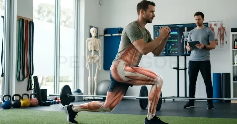
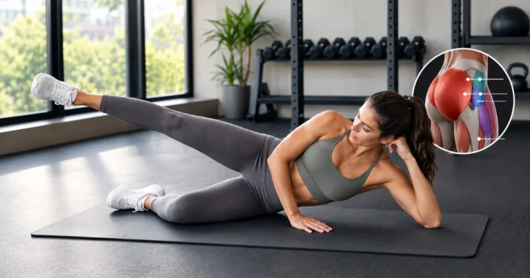
From lifting you out of a chair to powering you up a flight of stairs and cushioning your every landing, the quadriceps femoris muscle is the primary driving force of your lower body. This massive four-part group on the front of the thigh handles an immense daily workload that most of us take for granted. In this guide, we explore its intricate muscle anatomy, breaking down each individual head and the crucial tendon that connects them to the shinbone, while highlighting the key warning signs that signal it is time to seek a professional’s advice.
What the Quadriceps Femoris Group Includes
The quadriceps femoris muscle is a group of four muscles on the front of the thigh that work together to straighten the knee. The muscles of the quadriceps are the rectus femoris, vastus lateralis, vastus medialis, and vastus intermedius. Anatomy references describe the group as the most voluminous muscle mass in the body, so learning quadriceps muscle anatomy makes thigh and knee symptoms easier to understand.
Why It Is Called a Four-Headed Muscle
The quadriceps meaning comes from Latin: quadriceps translates as four-headed, and femoris means “of the femur,” the thigh bone. Each head starts at a different point on the pelvis or femur, yet all four fuse into a shared tendon above the knee. Some researchers also describe a small extra slip, the tensor of the vastus intermedius, but the classic four-head model remains the standard in the anatomical terminology used by clinicians in Europe and North America.
Anterior Compartment Position and Femoral Nerve Supply
The quadriceps lies in the anterior compartment of the thigh, and the femoral nerve powers all four heads. The thigh has three muscle compartments (anterior, medial, and posterior), and the front one also holds hip flexors such as the sartorius. Because the femoral nerve arises from spinal roots L2 to L4, a problem along that pathway can weaken several heads at once. Next, we look at the one head that behaves differently from the other three.
Rectus Femoris: The Only Two-Joint Quadriceps Head
The rectus femoris is the only quadriceps head that crosses both the hip and the knee. It runs straight down the center of the front thigh and is the most superficial of the four. Because it spans two joints, it helps extend the knee and also lifts the thigh at the hip. This double role shapes how it works, tires, and gets injured.
Straight and Reflected Heads From the Pelvis
The rectus femoris starts with two tendons on the pelvis, known as the straight head and the reflected head. The straight head attaches to the anterior inferior iliac spine, a small bony point at the front of the pelvis. The reflected head attaches to a groove just above the hip socket. The two tendons unite into one muscle belly that joins the shared quadriceps tendon above the kneecap.
Hip Flexion Contribution and Why It Fatigues Differently
The rectus femoris contributes to hip flexion, so it works harder than the other three heads whenever your hip and knee move together. Because it is the only head attached to the pelvis, it can lift the thigh as well as straighten the knee. You use this role in movements such as:
- Swinging the leg forward with each stride
- Lifting the knee to climb a high step
- Pedaling a bicycle, as commuters do daily in cities like Amsterdam and Copenhagen
Its fibers may also take turns working along the muscle, which anatomy reviews link to delayed fatigue. Because it is loaded at both joints, it tends to face more strain than the single-joint heads, a point we revisit in the injury section. Next, we compare the three vasti that attach directly to the femur.
Vastus Lateralis, Medialis and Intermedius Compared
The three vasti are single-joint muscles that attach to the femur and cross only the knee. Together these parts of the quad muscle supply most of the bulk and power among the muscles of the front thigh. Each has its own position, fiber direction, and role in guiding the kneecap, so we take them one at a time.
Vastus Lateralis and Lateral Thigh Bulk
The vastus lateralis sits on the outer thigh and, based on its internal architecture, is one of the strongest muscles in the lower limb. It begins on the greater trochanter, the gluteal tuberosity, and the lateral lip of the linea aspera, a ridge along the back of the femur. Its pull on the kneecap is slightly outward, so the inner structures of the knee must balance it.
Vastus Medialis Obliquus and Patellar Tracking
The vastus medialis obliquus (VMO) is the lower, more angled portion of the vastus medialis on the inner thigh. Its fibers run at roughly 40 to 55 degrees to the femur, which suits a role in guiding the kneecap along the groove it slides in. Electromyography studies, however, suggest that exercise cannot easily isolate the VMO from the rest of the quadriceps. Balanced strength across the whole thigh and hip is the safer goal.
Vastus Intermedius Beneath the Rectus Femoris
The vastus intermedius lies deep to the rectus femoris and rests directly on the front of the femoral shaft. You cannot see or feel it from the surface, yet imaging research suggests it makes up roughly 30 percent of quadriceps volume. Its deepest fibers form the articularis genus, a small slip that attaches to the knee joint capsule. Next, we follow all four heads down to their shared tendon at the kneecap.
The Extensor Mechanism: Quadriceps Tendon to Patella
The extensor mechanism is the chain of structures that carries force from the quadriceps femoris muscle to the shinbone. Doctors use this term because the chain works as one unit, and a break at any link can remove your ability to straighten the knee against gravity. The chain has five links, listed here in order from thigh to shin:
- The four quadriceps heads (muscle bellies)
- The quadriceps tendon above the kneecap
- The patella (kneecap)
- The patellar tendon below the kneecap
- The tibial tuberosity, the bony bump on the front of the shin
Patella as a Sesamoid Bone and Its Mechanical Advantage
The patella is a sesamoid bone, meaning it grows inside a tendon, and it is the largest sesamoid bone in the human body. By holding the tendon away from the knee’s axis of rotation, it lengthens the lever arm of the quadriceps, much like a pulley that redirects a rope. This mechanical advantage matters most in the last degrees of straightening. The patella also shields the tendon from rubbing against the femur.
Patellar Tendon and Tibial Tuberosity Insertion
The patellar tendon connects the lower tip of the patella to the tibial tuberosity on the front of the shin. Because it links two bones, many anatomists also call it the patellar ligament, and clinicians use both terms. You can feel it as a firm cord just below your kneecap when your knee is straight. All four heads send their force through this structure, so next we look at what that force accomplishes at the knee and hip.
What the Quadriceps Do at the Knee and Hip
The main job of the quadriceps is to extend the knee, and through the rectus femoris it also helps flex the hip. This dual quadriceps function, carried out by the quad muscles in the leg, supports standing, walking, and posture. Anatomy reviews describe the group as essential for climbing stairs and rising from a chair.
Knee Extension Force in Walking, Stairs and Squatting
Knee extension is the signature action of the quadriceps, and you rely on it in almost every upright activity. During walking it steadies the knee as your foot lands, and during stairs or squats it does much more of the work. Everyday examples of quadriceps strength at work include:
- Standing up from a chair or a low sofa
- Climbing the stairs of a walk-up apartment building, which are common in older European cities
- Squatting to lift a box or pick up a child
Because these tasks are so frequent, quadriceps weakness often shows up first as difficulty with stairs or low seats. Muscle mass and strength also tend to decline with age, a change called sarcopenia, so persistent or worsening difficulty deserves attention from a clinician.
Eccentric Control During Landing and Descent
Eccentric control means the quadriceps lengthen while producing force, acting as a brake on the knee. You use this braking action when you walk downstairs, hike down an Alpine trail, or land from a jump, because it provides smooth deceleration. Without it, the knee would fold too quickly, and long descents often leave the thighs sore the next day. Next, we show how to identify each head on a labeled image.
Labeled Quadriceps Diagram and Front Thigh Chart
A labeled quadriceps diagram shows the rectus femoris in the center of the front thigh, the vastus lateralis on the outer side, and the vastus medialis on the inner side. The vastus intermedius is hidden beneath the rectus femoris. The quad anatomy chart below summarizes the key facts about each head.
Head | Main origin | Insertion | Nerve | Main action |
Rectus femoris | Anterior inferior iliac spine and the groove above the hip socket | Patella via the quadriceps tendon, then tibial tuberosity | Femoral nerve | Extends the knee and flexes the hip |
Vastus lateralis | Greater trochanter, gluteal tuberosity, lateral lip of the linea aspera | Patella via the quadriceps tendon, then tibial tuberosity | Femoral nerve | Extends the knee |
Vastus medialis | Intertrochanteric line and medial lip of the linea aspera | Patella via the quadriceps tendon, then tibial tuberosity | Femoral nerve | Extends the knee and helps guide the kneecap |
Vastus intermedius | Front and lateral surfaces of the femoral shaft | Patella via the quadriceps tendon, then tibial tuberosity | Femoral nerve | Extends the knee, with deep fibers forming the articularis genus |
Identifying Each Head on an Anterior Thigh Image
You can identify each head on an anterior thigh image by working from the kneecap upward and then outward. Most labeled images use the same visual clues, so this sequence works for quad muscles labeled on textbook figures, clinic posters, and other quadriceps anatomy pictures:
- Find the patella at the bottom center, then trace the quadriceps tendon upward to the muscle bellies.
- Locate the rectus femoris, the long central muscle running down the middle of the thigh.
- Look to the outer side for the vastus lateralis, the widest and bulkiest head.
- Look to the inner side for the vastus medialis, which bulges lowest near the knee.
- Remember that the vastus intermedius is hidden and appears only when the rectus femoris is removed or drawn transparent.
Naming each head this way also helps you describe symptoms to a clinician, for example pain high in the central thigh versus pain just above the kneecap. That skill leads into the next topic, which is where problems most often arise.
Quadriceps Problems and Rehabilitation Context
Quadriceps problems usually fall into three groups: muscle strains, tendon problems, and pain around the kneecap. If you are dealing with one of them right now, please know that many respond well to an accurate diagnosis and gradual, supervised rehabilitation. This section offers general context only and cannot replace an examination by a licensed clinician.
Quadriceps Strain, Tendinopathy and Patellofemoral Pain
A quad strain is an overstretching or tearing of muscle fibers, and the rectus femoris is the head injured most often in any quadriceps injury. It typically happens during sprinting or kicking, when the muscle is stretched while it contracts. Tendinopathy is pain and wear in the tendon above the kneecap, and patellofemoral pain is aching around or behind the kneecap. The warning signs differ:
- Strain: sudden, sharp front-thigh pain during sprinting, kicking, or jumping
- Tendinopathy: pain just above the kneecap that builds with running or jumping
- Patellofemoral pain: a dull ache around the kneecap, worse on stairs or after long sitting
- Contusion (bruise): tenderness and swelling after a direct blow to the thigh
For patellofemoral pain, an international expert consensus recommends exercise therapy that combines hip and knee strengthening. Medicines matter too: the European Medicines Agency (EMA) advises stopping a fluoroquinolone antibiotic at the first sign of tendon pain or swelling and contacting your doctor. The risk of tendon damage is higher if you are over 60 or take a corticosteroid.
Post-Injury Muscle Atrophy and When to Seek Physiotherapy
Quadriceps atrophy is a loss of muscle size and strength that commonly follows knee injury, surgery, or long periods of reduced activity. Part of the weakness comes from the nervous system, which can limit muscle activation after joint injury, a process called arthrogenic muscle inhibition. Structured rehabilitation can help restore function.
Some symptoms need prompt medical attention rather than a routine physiotherapy appointment. Please contact a doctor or urgent care service without delay if you notice any of these signs, and call emergency services (112 in the European Union, 911 in the United States) after a severe injury:
- A pop or tearing sensation, followed by inability to straighten the knee or lift the leg
- A visible gap or dent just above the kneecap
- Severe, tense swelling of the thigh after a fall or blow
- Repeated giving way of the knee
- Thigh weakness that keeps worsening, especially with numbness
For milder, persistent problems, arrange an assessment if weakness or pain around the knee lasts more than a couple of weeks or limits daily activity. Physiotherapy (called physical therapy in the United States) is accessed differently across Europe, with some countries allowing direct booking and others requiring a referral, so your general practitioner is a reliable starting point. Finally, we explain how we sourced this guide.
Quadriceps Questions Readers Search For
These short answers cover three common questions readers ask about quad muscle anatomy. Each answer draws on the detail described earlier in this guide, so you can return to the matching section for more information whenever you need it. We keep each response brief and place the answer first.
What Are the Four Quad Muscles Called?
The four quad muscles are the rectus femoris, vastus lateralis, vastus medialis, and vastus intermedius. Together they form the quadriceps femoris and share one tendon at the kneecap, which is why they act as a single unit at the knee. This quick list shows where each one sits:
- Rectus femoris: center of the thigh, crossing both the hip and the knee
- Vastus lateralis: outer thigh
- Vastus medialis: inner thigh, including the VMO
- Vastus intermedius: deep, beneath the rectus femoris
Which Quadriceps Head Produces the Most Force?
No single head wins in every situation, but the vastus lateralis is most often credited with the greatest force-generating capacity. A cadaver study of muscle architecture ranked it among the strongest muscles of the lower limb. Ultrasound research in living adults, however, suggests that the deep vastus intermedius may best predict knee extension force. In practice, all four heads work together, and their share depends on knee angle and the task.
Where Exactly Is the Quad Muscle Located?
The quad muscle is located on the front of the thigh, between the hip and the knee. In everyday terms, it is the quad part of the leg, covering the front and sides of the femur, the thigh bone. You can feel it tighten when you straighten your knee from a seated position, and its shared tendon runs over the kneecap to the shin. Let us finish with the key points to remember.
The quadriceps femoris muscle is a four-headed group that straightens the knee, helps steady the kneecap, and, through the rectus femoris, assists the hip. In this guide, we covered its four heads, the extensor mechanism from tendon to shin, its role in daily movement, and the injuries and warning signs worth knowing. If you have questions or experiences to share, we welcome your comments below, and for any pain or weakness that worries you, please speak with a licensed clinician.