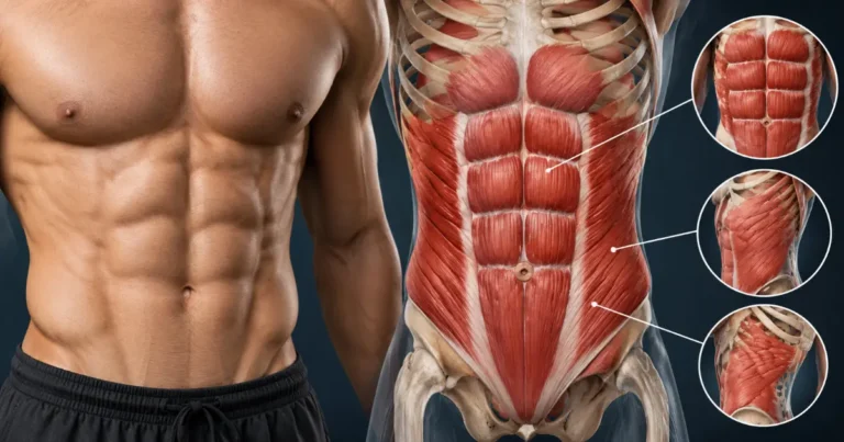
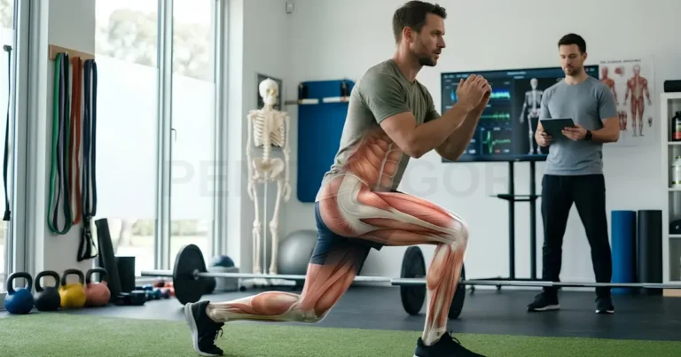
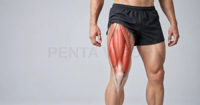
Have you ever wondered how your leg muscles power you through a busy day of walking and standing and why they sometimes unexpectedly ache or cramp? This comprehensive guide breaks down lower limb muscle anatomy from hip to foot, featuring labeled diagrams, nerve pathways, and blood vessels. We will also help you identify which symptoms are harmless and which require prompt medical attention.
How the Lower Limb Is Organized From Hip to Foot
The lower limb is organized from the hip to the foot into regions, and its muscles sit inside fascial compartments that share nerves and blood vessels. You can think of it as a building with separate floors, each with its own wiring and plumbing. Once you understand this layout, every muscle name becomes easier to place. For example, pain over the front of the shin points toward a different compartment than pain in the calf.
Regions of the Lower Limb in Anatomical Terminology
In anatomical terminology, the lower limb runs from the hip and gluteal region through the thigh, knee, leg, and ankle to the foot. Muscles are concentrated in the gluteal region, thigh, leg, and foot, while the knee and ankle are joints crossed by muscles from neighboring regions. Anatomists use the word leg only for the segment between the knee and the ankle, although most people use it for the whole limb. The checklist below shows where each muscle-bearing region begins and ends:
- Gluteal region: the buttock and hip area, home to the gluteal muscles that extend and stabilize the hip.
- Thigh: from the hip to the knee, containing the quadriceps, hamstrings, and adductors.
- Leg: from the knee to the ankle, containing the shin and calf muscles.
- Foot: from the ankle to the toes, containing small intrinsic muscles that support the arches.
Fascial Compartments as the Key to Understanding Leg Muscles
Fascial compartments are sections of muscle wrapped in tough connective tissue, and each compartment usually shares one nerve supply and one main artery. The thigh has three compartments (anterior, medial, and posterior), and the lower leg has four (anterior, lateral, superficial posterior, and deep posterior). Because the fascia is rigid, swelling inside a compartment raises pressure quickly, which is why a tight cast or a crush injury can threaten a calf. Next, we trace the nerves that supply each compartment.
Nerve Supply Across the Whole Lower Limb
The lower limb receives its nerve supply from the lumbosacral plexus, a network of nerves that emerges from the lower spine. Each nerve carries two kinds of signals: motor commands that make muscles contract, and sensory messages that report touch, pain, and position. When you understand this wiring, patterns such as numbness over the shin or difficulty lifting the foot start to make sense.
Lumbar and Sacral Plexus Contributions
The lumbar plexus and the sacral plexus are two nerve networks that together supply the entire lower limb. The lumbar plexus forms from the upper lumbar nerve roots and mainly serves the front and inner thigh. The sacral plexus forms from the lower lumbar and sacral roots and serves the buttock, the back of the thigh, the leg, and the foot. An irritated nerve root in the lower back can therefore cause symptoms far below the spine, such as calf pain or foot weakness.
Femoral, Obturator, Tibial and Common Fibular Territories
Four major nerves divide the lower limb into territories: the femoral, obturator, tibial, and common fibular nerves. The femoral and obturator nerves arise from the lumbar plexus, while the sciatic nerve, which also supplies the hamstrings, splits near the back of the knee into the tibial and common fibular nerves. Older texts use the term peroneal, but fibular is now the preferred name for the same nerve. The checklist below matches each nerve with the muscles it moves:
- Femoral nerve: the front of the thigh, including the quadriceps, which extend the knee.
- Obturator nerve: the inner thigh, including most adductors, which bring the leg toward the midline.
- Tibial nerve: the calf muscles, the deep posterior leg muscles, and the sole of the foot.
- Common fibular nerve: divides into the superficial branch (lateral leg muscles) and the deep branch (front of the shin and top of the foot).
These nerves rarely travel alone. Each runs beside an artery and a vein inside the same compartment, forming a neurovascular bundle, so an injury to one structure can affect its neighbors. A person who compresses the common fibular nerve at the outer knee, for example, may notice foot drop while walking. Now we follow the blood vessels that feed these muscles.
Arterial Supply and Venous Return in the Leg
Arteries carry oxygen-rich blood down the limb, and veins carry it back to the heart against gravity with help from the muscles themselves. Healthy circulation is what allows your leg muscles to keep working during a long walk. When either side of this system is disrupted, the muscles respond with pain, swelling, or fatigue.
Femoral, Popliteal and Tibial Arterial Pathway
Blood reaches the leg along one continuous pathway: the femoral artery in the thigh, the popliteal artery behind the knee, and the anterior and posterior tibial arteries below the knee. The femoral artery gives off the deep femoral artery, its largest branch, which supplies muscles of the thigh. The fibular artery, which usually arises from the posterior tibial artery, sends branches to the lateral compartment. You can feel this pathway as pulses at the groin, behind the knee, and beside the inner ankle.
Veins follow the arteries: deep veins run beside them, while the great and small saphenous veins run closer to the skin and drain into the deep system. When you walk, the soleus and gastrocnemius squeeze the deep veins, and one-way valves keep blood moving upward. Sitting still for hours, such as on a long flight, leaves this pump idle, which helps explain why the NHS lists long journeys as a time of higher clot risk. With the supply lines mapped, we can meet the muscles, starting with the thigh.
Upper Leg Muscle Compartments Summarized
The upper leg, or thigh, contains three muscle compartments, and each one has a distinct main action at the hip or knee. The front compartment mostly extends the knee, the back compartment flexes the knee and extends the hip, and the inner compartment draws the thigh toward the midline. The upper front leg muscles do most of the heavy lifting when you rise from a chair, with help from the other muscles on the upper leg.
Anterior, Posterior and Medial Thigh Compartments Compared
The anterior, posterior, and medial thigh compartments differ in their muscles, their movements, and their nerve supply. The adductor magnus is a notable exception because it receives supply from both the obturator and sciatic nerves. The table below compares the three compartments at a glance:
Compartment | Main muscles | Main action | Nerve supply |
Anterior | Quadriceps (four parts), sartorius | Extends the knee, flexes the hip | Femoral nerve |
Medial | Adductor longus, brevis, and magnus, gracilis | Draws the thigh toward the midline | Obturator nerve |
Posterior | Biceps femoris, semitendinosus, semimembranosus | Bends the knee, extends the hip | Sciatic nerve |
A hamstring strain during sprinting, for example, involves the posterior compartment, while a groin strain typically involves the adductors. Naming the compartment helps you describe symptoms accurately to a clinician. Below the knee the organization changes, so we turn to the lower leg next.
Lower Leg Muscle Compartments Summarized
The lower leg has four muscle compartments: anterior, lateral, superficial posterior, and deep posterior. Some texts count three by grouping both posterior layers together. These lower muscles of the leg lift the foot, turn it outward, and push it off the ground, and the tibia, the fibula, and a membrane between them form the walls of the compartments.
Anterior, Lateral and Posterior Shank Compartments Compared
The anterior compartment lifts the foot, the lateral compartment turns it outward, and the two posterior compartments push it downward and flex the toes. Anatomists sometimes call the segment between knee and ankle the shank. Each compartment has its own nerve: the deep fibular nerve serves the front, the superficial fibular nerve serves the outer side, and the tibial nerve serves the back. The table below compares all four compartments:
Compartment | Main muscles | Main action | Nerve supply |
Anterior | Tibialis anterior, extensor hallucis longus, extensor digitorum longus, fibularis tertius | Lifts the foot and extends the toes | Deep fibular nerve |
Lateral | Fibularis longus, fibularis brevis | Turns the foot outward | Superficial fibular nerve |
Superficial posterior | Gastrocnemius, soleus, plantaris | Points the foot downward | Tibial nerve |
Deep posterior | Tibialis posterior, flexor digitorum longus, flexor hallucis longus, popliteus | Supports the arch, flexes the toes, and helps start knee bending | Tibial nerve |
The calf shows why this matters. The gastrocnemius forms the visible bulge, and the soleus lies beneath it, so both can tire after a long hill walk. Pain over the front of the shin, by contrast, may involve the anterior compartment. Beyond the ankle, another set of small muscles takes over inside the foot itself.
Intrinsic Foot Muscles and the Arch System
Intrinsic foot muscles are small muscles that begin and end inside the foot, and together with ligaments and the plantar fascia they support the arches. Counting the small lumbrical and interosseous muscles individually, each foot has about 20 of them. Together with the arch, they help the foot absorb impact and then stiffen for push-off.
Plantar Muscle Layers and Their Role in Gait
The plantar muscles are arranged in four layers, from the most superficial layer beneath the plantar fascia to the deepest layer beside the metatarsal bones. Early in stance the arch flexes to absorb impact, and later it stiffens through the windlass mechanism, in which lifting the big toe tightens the plantar fascia and raises the arch. Researchers still study how much active muscle work adds to this passive tissue support. The checklist below names the muscles in each layer:
- Layer 1 (most superficial): abductor hallucis, flexor digitorum brevis, and abductor digiti minimi.
- Layer 2: quadratus plantae and the four lumbricals.
- Layer 3: flexor hallucis brevis, adductor hallucis, and flexor digiti minimi brevis.
- Layer 4 (deepest): the dorsal and plantar interossei.
For example, heel pain that is worst with the first steps after rest is a common pattern of plantar fasciitis, an overuse condition of the fascia that runs from the heel to the toes. If you notice this pattern, a clinician can confirm the cause and suggest treatment. With every region described, we can now place these muscles on a labeled diagram.
Labeled Leg Muscle Diagram: Front, Back and Side Views
A labeled leg muscle diagram shows the major muscles from three viewpoints, and each view highlights a different set of structures. The front view of a leg muscle chart emphasizes the quadriceps, sartorius, and tibialis anterior. The back view shows the gluteals, hamstrings, and calf, and the side view shows the iliotibial band and the fibularis muscles. Any good picture of leg muscles should label all three views.
Matching Chart Labels to Palpable Landmarks
Matching chart labels to palpable landmarks means finding each muscle on your own body using bones and tendons you can feel through the skin. This turns a flat chart into a working map, a skill that anatomy students and physical therapists use daily. Use the steps below to practice leg surface anatomy with the chart beside you. Stop if anything hurts, and remember that these are learning aids rather than diagnostic tests:
- Find the bony point at the front of the pelvis (anterior superior iliac spine), where the sartorius begins.
- Locate the kneecap, then trace upward to the quadriceps tendon and downward to the patellar tendon.
- Feel the sharp front edge of the shin bone (tibia); the tibialis anterior lies just outside it.
- Find the rounded head of the fibula at the outer knee, where the fibularis muscles begin.
- Follow the calf down to the Achilles tendon, which attaches to the heel bone.
- Bend the knee gently while touching the back of the thigh to feel the hamstring tendons behind the knee.
For example, if you feel tightness at the outer knee, noting whether it sits near the head of the fibula or higher along the thigh helps a clinician narrow down which structures are involved. Precise descriptions save time during an appointment. With the map in hand, we can now see how these muscles cooperate in motion.
How Leg Muscles Coordinate During Walking and Running
During walking and running, leg muscles coordinate in a repeating gait cycle that alternates between a weight-bearing stance phase and a swing phase. In walking, about 60% of the cycle is spent in stance and about 40% in swing. Running keeps the same alternating pattern, with a shorter stance phase and a brief moment when both feet are off the ground.
Stance Phase, Swing Phase and Muscle Activation Order
The stance phase begins at heel contact, when the tibialis anterior and quadriceps work eccentrically, meaning they lengthen while contracting, to control the landing. In mid and late stance, the gluteus medius steadies the pelvis while the gastrocnemius and soleus control the shin and then drive the push-off. In swing, the hip flexors and tibialis anterior lift the limb and clear the toes, and the hamstrings slow the leg before the next heel contact. If the tibialis anterior is weak, the toes may catch and the foot may slap down.
Gait Cycle Table: Phase, Active Muscles and Function
The table below summarizes the main phases of walking, the gait muscles most active in each, and the job they perform. Timing and intensity vary with walking speed, footwear, age, and health conditions, so please treat it as a general reference rather than an exact recording.
Phase | Main active muscles | Function |
Initial contact | Tibialis anterior, quadriceps, hamstrings, gluteus maximus | Controls the landing and steadies the ankle and knee |
Loading response | Quadriceps, tibialis anterior, gluteus medius, adductor magnus | Absorbs shock and stabilizes the pelvis |
Midstance | Gastrocnemius, soleus, gluteus medius | Controls shin advance and keeps the pelvis level |
Terminal stance | Gastrocnemius, soleus | Lifts the heel and drives push-off |
Pre-swing | Iliopsoas, rectus femoris | Prepares the limb to leave the ground |
Initial and mid swing | Iliopsoas, tibialis anterior | Swings the limb forward and clears the toes |
Terminal swing | Hamstrings, quadriceps, tibialis anterior | Slows the limb and prepares for heel contact |
You can notice this coordination when you walk downhill. The quadriceps work eccentrically to brake your body weight, a pattern many people feel as sore thighs the next day. When coordination or blood supply falters, however, symptoms can appear that are not ordinary soreness. The next section explains which symptoms deserve clinical attention.
Leg Muscle Symptoms That Need Clinical Attention
Many leg muscle aches after unusual activity settle within days, but a few patterns need prompt medical assessment. Because leg pain can come from muscles, nerves, or blood vessels, only a clinician can tell them apart. We share the patterns below so you can act early, not so you can diagnose yourself.
Compartment Syndrome, Claudication and Persistent Cramp
Compartment syndrome, intermittent claudication, and persistent cramp are three distinct patterns of leg pain that call for medical advice. Compartment syndrome is a rise in pressure inside a muscle that restricts blood flow, and the NHS treats sudden, severe pain as an emergency. Intermittent claudication is cramp-like leg pain that starts with walking and eases within minutes of rest, and it can signal peripheral arterial disease. Persistent cramp is a painful tightening that keeps returning and can have several causes, so it deserves a proper check.
Some medicines can also affect muscles and tendons. The European Medicines Agency (EMA) recommends that patients be advised to stop a fluoroquinolone antibiotic at the first sign of muscle pain, weakness, or tendon problems. A European Atherosclerosis Society panel reports muscle symptoms in roughly 7% to 29% of statin users in observational studies, so please discuss symptoms with your prescriber before changing a dose. The checklist below groups warning signs by urgency:
- Emergency (call your local emergency number): sudden, severe leg pain that does not ease, or a tight, bulging muscle after an injury or cast.
- Emergency: a leg or foot that suddenly turns pale, blue, cold, or numb.
- Emergency: calf pain and swelling together with chest pain or breathlessness, which can signal a blood clot that has reached the lungs.
- Emergency: new leg weakness together with numbness between the thighs or around the buttocks, or new bladder or bowel control problems, which can signal cauda equina syndrome.
- Urgent (same-day advice): throbbing pain, swelling, warmth, and redness in one calf or thigh, which can signal deep vein thrombosis.
- Urgent (same-day advice): new weakness in the leg or a foot that drags when you walk.
- Book an appointment: cramp-like pain that starts with walking and eases with rest.
- Book an appointment: pain, numbness, or swelling that returns with exercise and settles with rest.
- Book an appointment: cramps that keep returning, muscle wasting, or new muscle pain after starting a medicine.
For claudication, the 2024 European Society of Cardiology guidelines recommend supervised exercise training for symptomatic peripheral arterial disease, which shows how much can be done once the cause is identified. When you see your family doctor, bring a list of your medicines and a note of when the pain starts and what relieves it, and ask your pharmacist about the package leaflet if a medicine may be involved. Next, we explain how this guide was researched.
Research Standards Applied to This Lower Limb Guide
For this guide, we applied four research standards: authoritative sources, current terminology, European relevance, and transparent limits. We prioritize clinical guidelines from bodies such as the ESC and the NHS, regulatory notices from the EMA, and peer-reviewed studies. For basic anatomy, we use clinical reference texts such as StatPearls and established medical education sites. We use the current name fibular, noting the older term peroneal at first mention, and we say so when anatomy counts or names vary between textbooks.
This guide is for education and does not replace personal medical advice. Articles on Penta Vigor are not a substitute for a medical consultation, and nothing here should delay you from seeking care. We gathered the sources listed at the end of this guide in September 2026, and if you spot an error or a newer guideline, please tell us so we can correct the guide. The numbered list follows the order in which each source first appears.
Common Leg Muscle Inquiries
Below we answer three questions we hear most often about the anatomy of the lower limb. Anatomy sources do not always agree on exact counts, so we explain the reasoning behind each answer. Use these short answers as a starting point, and return to the earlier sections for details.
How Many Muscles Are in the Leg?
There is no single agreed number, but each lower limb contains more than 40 muscles when the hip, thigh, leg, and foot are all counted. Lower figures appear in some sources because they count the four parts of the quadriceps as one muscle. The foot alone holds about 20 intrinsic muscles. The number you see depends on where the author draws the boundary of the leg.
What Is the Largest Muscle in the Leg?
The gluteus maximus is the largest muscle in the body, although anatomists place it in the gluteal region rather than in the leg proper. Within the thigh, the quadriceps is the largest muscle group, and the vastus lateralis is its largest part. The sartorius, which crosses the front of the thigh, is often described as the longest muscle in the body. So the answer depends on whether you mean largest by mass, group size, or length.
Which Leg Muscles Work Hardest in Daily Movement?
The soleus, quadriceps, and gluteal muscles are among the muscles that work hardest in daily life, because they resist gravity whenever you stand, walk, or climb stairs. The soleus is rich in slow-twitch, fatigue-resistant fibers, about 70% in one classic autopsy study, which suits its constant postural role. It also contributes to the calf muscle pump that returns blood from the leg. Researchers measure effort in different ways, so rankings vary, but regular movement breaks during long trips or desk work give these muscles a chance to work.
In summary, your leg muscles are organized in compartments that share nerves and blood vessels, from the gluteal region through the thigh and lower leg to the foot, and they coordinate in a precise sequence with every step. Understanding this structure helps you describe symptoms clearly and recognize warning signs that need prompt care. If you have questions or suggestions for topics we should cover next, please share them, and speak with a licensed clinician about any symptom that worries you.